
44 Introduction to Memory
Chapter Outline
- How Memory Functions
- Memory and the Brain
- Problems with Memory
- Ways to Enhance Memory
Most of us would agree that memory is one of our most valuable assets. Our lives would be empty if we couldn’t store new information and our ability to access past experiences is a big part of who we are. Our nervous systems are bombarded with information and the brain continually sorts, organizes, stores, and retrieves throughout our lifetimes. Unlike physical storage devices, the brain can’t add another filing cabinet or hard drive. Instead, there are many different neural systems that overlap and influence each other to create and retrieve memories. Scientists have been able to define distinct memory processes associated with activity in specific brain regions.
Studying the role of the brain in memory requires isolating a specific region from the larger network of cognitive systems. One approach is by intentionally damaging a brain area, called ablation or lesioning, and then looking at the effects on memory. If the ablation procedure impairs performance on a memory task, it suggests that the damaged part of the brain was involved. Ablation studies in non-human animal models allow researchers to control the location and timing of damage but purposely damaging the brain of human participants for research isn’t possible. Therefore, these types of questions in humans usually rely on individuals with damage to the brain from injury or disease. In other words, researchers work “retrospectively” by looking back at the consequences of an event that has happened in the past on current memory.
People with rare brain injuries can provide unique information about the nature of memory by examining the abilities that they lost. One famous case study of memory comes from a man named Henry Molaison, known as patient HM, and his collaboration with researcher, Brenda Milner. After receiving a bilateral medial temporal lobectomy to treat seizures, HM experienced severe memory loss, providing a unique opportunity for Milner to study how the structures he lost impacted his ability to create new memories and learn new skills. The groundbreaking findings from this collaboration would later become the foundation for the fields of cognitive neuroscience and clinical neuropsychology.
By the early 1950’s, removal of large areas of cortex, called lobectomies, were used to treat many different neurological conditions. In 1955, Dr. William Scoville operated on a 27-year old assembly line worker, Henry Molaison, to control his seizures. His anticonvulsant were ineffective, so Scoville proposed a temporal lobectomy, removal of the anterior hippocampus, parahippocampal gyrus, and amygdala. Previous patients usually didn’t experience severe symptoms aside from some mild memory impairment, but post-surgery HM experienced severe and unexpected loss of memory (Milner, 2005). He could not recognize the hospital staff or Dr. Scoville himself, whom he’d known for many years. After his procedure, he forgot everything moment to moment and could not create any new memories about what happened each day at the hospital, not even the way to the bathroom. (Milner, 2005; Scoville 1954).
Despite this severe memory impairment, HM still had his early-life memories and retained his capacity for speech, social behaviour, and emotional responses. He also scored higher on the Weschler IQ test than he had before the surgery, probably because his seizures had stopped. At any given moment, HM described his world as being “clear” but said he felt like he was always waking up from a dream (Milner, 2005). After his surgery, HM was moved to a new house only a few blocks away from where his family had been living and it took about five years to navigate the inside of his home (Milner, 1970).
After observing this severe memory loss in HM following his surgery, Scoville invited Brenda Milner, a researcher from the Montreal Neurological Institute with expertise in the function of the medial temporal lobes (Milner, 2005). In order to test HM’s capacity for short term memory, Milner adapted a spatial task that asked participants to discover the correct route through a digital maze along a series of “stepping stones”. Using trial and error, the participant selects a stone along the path and is given feedback if they are correct. If not, the participant is sent back to the beginning of the maze to try again. Most people can remember enough correct steps to make it through the maze after about 20 trials, but HM failed to remember anything after 215 attempts (Milner, 1966; Milner 2005). Milner later trained him on a much shorter version of the maze with only six choice points, which did not exceed his immediate memory span. Although it took 155 trials and a lot of effort, HM was eventually able to complete three errorless runs through the maze, and even remembered some of the correct pathways two years later (Milner et al., 1968; Milner, 2005).
These findings from the digital maze study revealed two key points. First, HM was able to remember the pathway once the stepping stones were reduced to six, showing that he had some capacity for immediate memory. Second, he kept the memories that he was able to create for years. We can make two conclusions from these findings. First, his immediate memory functioned normally even though his ability to form longer-term memories was impaired, and second, forming memories and keeping memories could require different processes. Because HM was missing his medial temporal lobes, we can assume that this area of the brain is important for forming memories, but not necessarily storing them. This is a simple example of how a retrospective ablation study order to make links between different brain areas and the behaviours they might control.
Milner used many different tests and techniques with HM and their collaboration became one of the most influential in neuroscience since it helped link the medial temporal lobe to specific memory function and aided in distinguishing different types of memory. By observing what HM was not able to do, Milner and her team could determine what the medial temporal lobe did control. It was obvious from the beginning that HM could not create any new memories of events in his life, and limited memory of new facts and information (Tulving, 1972). Over the years, Milner and other researchers noticed that HM struggled to gain new facts that had to do with changing technology and that he had learned fewer words than healthy people his own age. Nevertheless, Milner reported that he was somewhat aware of the Kennedy assassination occurring and of space travel (Gabrieli et al., 1988; Milner, 2005).
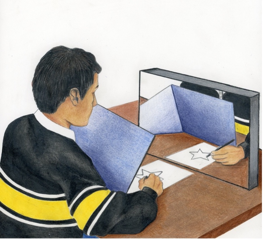
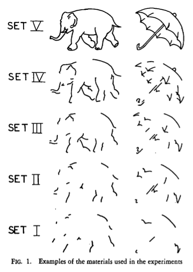
Milner was also able to determine which parts of memory were not controlled by the medial temporal lobes through her discovery of HM’s capacity for implicit learning, the act of gaining new skills without conscious awareness. First, he completed a motor task where he had to copy a drawing by only looking at a reflection of his hand through a mirror (Figure M.1). After 30 trials over three days, he had no conscious awareness of doing the task before, but his performance improved significantly (Corkin 1968; Milner, 1970; Milner, 2005). For the second task, HM had to identify an object shown as multiple incomplete line drawings, each differing in its level of completion (Figure M.2). It was nearly impossible for him to identify objects from the most incomplete drawings, but with practice he needed fewer cues to name the items (Gollin, 1960; Milner, 1970; Milner 2005). These studies showed that that the medial temporal lobes were not required for these types of memory tasks.
Thanks to Brenda Milner’s work, we’ve learned a lot from HM himself. After dedicating 30 years of his life to the research of memory, Henry Molaison also donated his brain for further study, upon his death in 2008 (New York times, 2009). The 53-hour brain dissection at the San Diego Brain Observatory, was streamed live to hundreds of thousands of viewers (Brain Observatory, 2009). The result of this procedure was 2041 high resolution images of ultra-thin brain sections, compiled in a virtual brain atlas available to researchers online (Annese et al., 2014). Despite his contributions to science, it was only after his death that HM became known by his full name, Henry Molaison, rather than his initials. His collaboration with Brenda Milner’s demonstrated there are many paths to learning and together they provided a deeper understanding of how our own minds work.
LINK TO LEARNING
